

Origins and the turning point
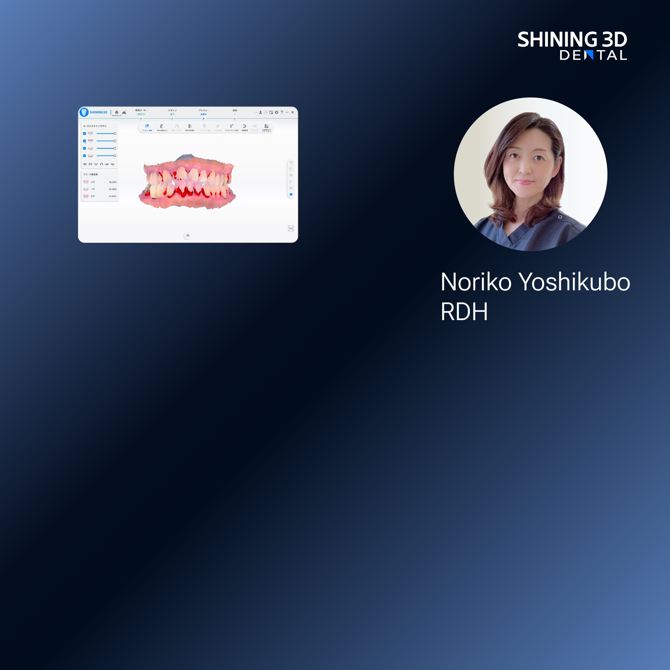
Dentistry kept careful records long before pixels arrived; impressions were plastered, adjusted, and eventually archived. The story shifted when chairside capture moved from curiosity to commonplace—now the intraoral scanner replaces repeated alginate trials and yields a reproducible digital starting point. A turning point is visible at major gatherings, such as recent editions of the International Dental Show in Cologne, where demonstrations of the dental intraoral scanner confirmed that clinicians expect predictable digital captures before design begins.
Stepwise transformation: how the workflow matured
The earliest step remains capture, but the method changed. The digital workflow now begins with high-resolution scanning or careful photograph-based records. Next comes data alignment: meshes are cleaned, indexed and keyed to occlusion. CAD receives those files and translates anatomy into prosthetic form; designers choose flange height, tooth selection, and retention mechanics with parametric controls. Prototyping follows—either rapid-milled try-ins or printed templates for clinical verification—then final fabrication by milling or additive processes and careful post-processing before clinical delivery. Each stage replaced a manual check with a measurable one: repeatable scans, versioned CAD files, and traceable milling parameters.
Why this evolution matters: concrete gains and trade-offs
Accuracy improved where analogue variance once dominated: repeatable digital capture cuts remakes. Time shifted from technician guesswork to designer verification, but new bottlenecks emerged—file-transfer errors, incompatible libraries, and clinician unfamiliarity with digital margins. Costs redistributed: investments moved from consumables toward scanners and software subscriptions. Those who mastered the digital chain reduced chairside adjustments; those who treated it as a single-device swap found more problems than solutions.

Alternatives tested along the way
Practices did not convert uniformly. Some adopted hybrid paths—analog impressions scanned later in the lab. Others leaned fully digital, while a few used desktop scanners rather than direct intraoral capture. Conventional methods still outperform digital on certain soft-tissue nuances and in cases with extreme resorption. Hybrid workflows offer a middle ground: faster turnaround than fully analogue routes and fewer surprises than an uncalibrated digital pipeline.
Common mistakes and pragmatic fixes
Overconfidence in software is a frequent error. Practitioners assume a perfect scan will always yield a perfect fit; missing soft-tissue detail or failing to register occlusion remains a real cause of remakes. A second mistake is ignoring milling and material limits: a design that looks fine on screen can fracture if wall thickness is below the material minimum. Fixes are straightforward—verify scans with a physical check, use validated material libraries, and run a prototype try-in before final fabrication. Train teams on a single, repeatable protocol rather than switching tools mid-process.
What to weigh when choosing equipment and partners
Decide by use-case. For in-clinic rapid turnaround, favor scanners and mills that integrate with your CAD system and offer local support. For complex esthetic cases, value open-file compatibility and a lab partner versed in prosthetic nuances. Evaluate uptime and service history; machines with predictable maintenance windows yield more consistent patient outcomes than those chosen solely on specs.
Concluding synthesis
The digital denture workflow reads like a sequence of chapters: capture, translate, verify, fabricate, deliver. Each chapter grew from practical problems: unreliable impressions, opaque lab steps, and unpredictable fit. Understanding that lineage clarifies which choices reduce risk and which introduce it. The present advantage rests on systems that connect clinical capture to material-aware fabrication and experienced technicians who respect both data and soft tissue. That linkage is where SHINING 3D DENTAL fits naturally into practice narratives—tools and protocols designed to bridge historical technique with controlled digital steps, so clinicians move from hopeful trial to reliable prosthesis with fewer surprises.
